
By Michael J. North

NLM #2295005
This year we commemorate the 500th anniversary of the birth of Andreas Vesalius (1514–1564) who is best known for changing how we do medical research with his groundbreaking book, De Humani Corporis Fabrica Libri Septem (Seven Chapters on the Structure of the Human Body), published in 1543 and generally known as De Fabrica.
One of the great mysteries surrounding Andreas Vesalius’s De Fabrica is who illustrated this beautiful and important book, which not only began a new era in medical research in which physicians made their own observations rather than relying on texts by the ancients, but also raised the importance of illustration to medical education and ushered in a new phase in medical publishing and the arts.
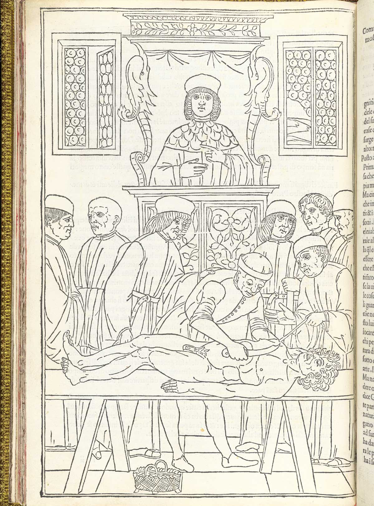
While at his post as professor of anatomy and surgery at the University of Padua, Andreas Vesalius spent much of 1542 preparing his magnum opus, the De Fabrica, performing dissections (sometimes in his home) and working with an artist (or more likely several artists) to create the woodcut illustrations for which the book is so well known. Vesalius was exacting in his methods of dissection and investigation of the cadaver. He would perform a dissection and position the cadaver so that important structures were highlighted and visible; he even describes in De Fabrica his use of nooses and cords to hold the cadavers in place. It is unknown, however, who the artist or artists were; it is likely that some of the images were first sketched by Vesalius himself and a final draft created by a trained artist (these are not the work of an amateur). Later, a craftsman, most likely in Venice, carved the images in relief onto blocks of pear wood. Correspondence between Vesalius and his publisher Johannes Oporinus describe the packing and transport of the woodblocks across the Alps from Venice to Basel, but no mention is made of the artists who created them.
One theory that held sway for many years was that the artist was Jan Steven van Calcar (1499-ca. 1546), a Flemish painter who was a student of Titian in Venice. A strong piece of evidence is that it is known that Calcar was the artist for Vesalius’s first illustrated anatomy, Tabulae Anatomicae, which came out in 1538. Many years later, Giorgio Vasari stated in the second edition of his noted Lives of the Most Famous Painters in 1568 that Calcar was responsible for the figures in Vesalius’s book, however, many believe that this is only a reference to Tabulae Anatomicae and not De Fabrica. Calcar died in 1546 or 1547, only a few years after De Fabrica was published, and he does not seem to have been described as Vesalius’s artist until Vasari’s book came out over 20 years later.
After the publication of De Fabrica, most of the woodblocks were stored and then re-used in the second edition of 1555, and many were used again and again for over a century. Almost unbelievably, Archibald Malloch, the Librarian at The New York Academy of Medicine in the 1930s, discovered that the woodblocks still survived at the Library of the University of Munich, and an edition of them was reprinted in 1934; sadly, they were destroyed during World War II.


Outstanding among the woodcut illustrations are the skeleton and muscle figures and the title page. The muscle figures are noted for their beauty and detail—the muscles had never been depicted in print in a way which allowed such close examination—and they are indexed by letters (some in Greek) pointing to different structures and described in the text of adjoining pages. The rural landscape in the background of the figures, which have been identified as the Euganean Hills outside Padua, form a continuous panorama of the area. The title page of De Fabrica is by itself considered one of the most famous woodcut illustrations of the 16th century because of its intricacy. Front and center in the elaborate scene is Vesalius himself with his hands on a female cadaver in an anatomical theater, instead of on a high chair reading a Galenic text while a surgeon or student performed the cutting. Characters in the title-page scene appear to include students, onlookers from the public, a skeleton, contemporary sages, and even what may be an ancient physician. Also depicted are a dog with a human foot and a rhesus monkey, most likely to symbolize the fact that Galen drew most of his anatomical knowledge from the dissection of these animals instead of humans.
The National Library of Medicine has scanned and made available over 40 pages of these woodcut images at high resolution, and many of them are described in the Library’s Turning the Pages project featuring the De Fabrica. The National Library of Medicine has a large collection of works by and about Andreas Vesalius and his groundbreaking approach. To learn more about them, please feel free to contact us at NLM Customer Support.
This article is the third in a series to commemorate the 500th anniversary of the birth of the great anatomist Andreas Vesalius, born on December 31, 1514.
 Michael J. North is the Head of Rare Books and Early Manuscripts in the History of Medicine Division at the National Library of Medicine.
De Fabrica was the Grey’s anatomy of its day, a truly stunning advancement for the practice of medicine
It certainly was. We hope you’re enjoying this series about it’s author.